Thymosin β4’s Protective Role in Myocardial Infarction and Cardiac Fibrosis
“Inflammation is a critical factor in the development and progression of myocardial infarction and cardiac fibrosis. Thymosin β4 (Tβ4) alleviates the disease process via protective antioxidant and anti-inflammatory mechanisms. Although Tβ4 has been shown to have a protective effect in myocardial infarction, its impact on cardiac fibrosis has not been well reported. In this study, we evaluated the influence of exogenous Tβ4 on myocardial infarction and cardiac fibrosis and explored the possible underlying mechanism… Tβ4 was shown to be significantly elevated in mice AMI cardiac tissues. In mice, AAV-Tβ4 induced exogenous expression of Tβ4 significantly reduced oxidative damage, inflammation, cardiac dysfunction, and fibrosis… AAV-Tβ4 induced expression of Tβ4 reduced inflammation, heart damage, and eventual fibrosis in vivo. Tβ4 helped to reduce oxidative stress, promote mitophagy, and alleviate inflammation and fibrosis. Exogenous supplementation of Tβ4 might be a promising therapeutic agent for treating myocardial infarction as well as cardiac fibrosis.” (3)
The Role of Thymosin β4 in Myocardial Fibrosis and Inflammation
“Myocardial fibrosis is characterized by a significant accumulation of extracellular matrix (ECM) in the myocardium… At the site of acute myocardial infarction (AMI), the sudden loss of a large number of cardiomyocytes triggers an inflammatory reaction, ultimately leading to the replacement of necrotic myocardium with a collagen-based network… Therapy for myocardial infarction and cardiac fibrosis frequently employs antioxidant, anti-inflammatory, and antifibrotic medicines. However, no therapeutic strategy has been developed that ensures damaged tissue reversal… Thymosin 4 (Tβ4) is a 43-amino acid protein that belongs to the β-thymosin family, which is highly conserved… Tβ4 has been associated with wound healing, inflammation, fibrosis, and tissue regeneration, with recent studies suggesting that Tβ4 can help prevent inflammation and fibrosis in the eye, skin, lung, and liver… Tβ4 is a potent protective factor that can protect against myocyte damage, promote myocyte regeneration, and inhibit heart inflammation… Therefore, we propose that Tβ4 might have an antifibrotic function in the heart. Tβ4 inhibits myofibroblast growth and TGF-β1-induced activation. Development of myofibroblast cells treated by Tβ4 (75-150 nM, 12 h).
In the present study, we first provided substantial evidence for the increased expression of Tβ4 in murine models of ligation-induced AMI and cardiac fibrosis. The role of Tβ4 in alleviating hepatic, renal, and cardiac injury and fibrosis has been confirmed in recent studies. The increased production of local Tβ4 in mice is an adaptive response to heart injury, but this increased expression of endogenous Tβ4 might not be sufficient to alleviate heart injury and fibrosis. In the present study, we observed the effects of adeno-associated virus-mediated Tβ4 ectopic expression on ligation-induced AMI and subsequent cardiac fibrosis. Our findings indicated a protective role of Tβ4 against oxidant damage and inflammasome activity, thereby alleviating myocardial infarction and cardiac fibrosis.
Previous studies have revealed the role of oxidative stress in the pathogenesis of cardiac inflammatory responses; the generation of mitochondrial ROS is crucial for NLRP3 inflammasome activation, leading to the release of IL-1β. Under inflammatory conditions, infiltrated and activated inflammatory cells, such as neutrophils, monocytes/macrophages, and eosinophils, can generate ROS via multiple enzymes and reaction pathways, including nicotinamide adenine dinucleotide phosphate oxidases, eosinophil peroxidase, and especially MPO. MPO catalyzes the formation of potent cytotoxic oxidants. The relationship between myocytes, oxidative stress, and inflammation then creates a vicious cycle. Here, our in vitro data demonstrated that ROS promoted inflammation in myocytes, which is consistent with previous findings that myocyte injury leads to the secretion of IL-1β. We also found that H2O2 treatment induced ROS generation in myocytes, thereby leading to the activation of the NLRP3 inflammasome, but this effect was alleviated by NAC, an antioxidant.
Although debates exist, inflammation is believed to contribute to the pathological progress of AMI and cardiac fibrosis, especially during the initial period. Infiltration of leukocytes into the heart leads to myocyte dysfunction and tissue damage, which trigger fibrogenic progression. Moreover, infiltrated leukocytes and damaged tissue cells can release proinflammatory cytokines, such as IL-1β, TNF-α, and IL-6, which also exert fibrogenic effects. Similarly, our present study demonstrated that Tβ4 alleviated ligation-induced heart inflammation as well as the production of profibrotic cytokines, suggesting that the anti-inflammatory potency of Tβ4 contributed to its antifibrotic effect.
Excessive ROS production is a hallmark of many diseases; dysfunctional mitochondria have been implicated in these disorders, acting as both a source and a target of ROS. Mitophagy is a kind of selective autophagy in which damaged or undesired mitochondria are degraded. In this study, myocyte mitophagy was discovered to be impaired, and NAC was found to alleviate this condition. Furthermore, we discovered that FCCP, a mitophagy inducer, inhibited H2O2-induced IL-1β production in myocytes, whereas oligomycin, a mitophagy inhibitor, enhanced production. Defective mitophagy leads to the buildup of damaged ROS-generating mitochondria and the activation of the NLRP3 inflammasome. Our data revealed for the first time that ROS promotes inflammation via mitophagy inhibition in myocytes.
Tβ4 has been shown to have antioxidant and anti-inflammatory properties. AAV-Tβ4 induced exogenous expression of Tβ4, which effectively reduced infarction-induced increases in mouse cardiac MPO activity, MDA levels, and proinflammatory cytokines in vivo, according to our findings. Inflammation is thought to have a role in the initial etiology of myocardial infarction and cardiac fibrosis; myocyte dysfunction and subsequent inflammation initiate the fibrogenic process, which results in matrix deposition and heart remodeling. Our data demonstrated that exogenous Tβ4 reduced infarction-induced myocardial damage and cardiac fibrosis in mice and decreased the fibrogenic process in myofibroblasts. In addition to protecting the heart from oxidative injury, our study demonstrated that Tβ4 promoted myocyte growth but attenuated myofibroblast growth. Tβ4 is a key factor in cardiac development, growth, disease, epicardial integrity, and blood vessel formation and has cardio-protective properties. The proliferation-promoting effect of Tβ4 might facilitate the repair of damaged myocytes in a manner that avoids aberrant repair leading to fibrosis. In contrast to promoting the proliferation of myocytes, Tβ4 suppressed the growth of myofibroblasts, which should inhibit the accumulation of ECM and further alleviate fibrogenesis.” (3)
Thymosin β4’s Impact on Mitophagy and Oxidative Stress in Myocardial Infarction and Cardiac Fibrosis
“Autophagy is a highly conserved mechanism that maintains homeostasis by catabolizing cytoplasmic components, such as defective proteins and organelles… Autophagy contributes to the end of NLRP3 inflammasome activation by targeting reactive oxygen species ROS-producing mitochondria. The process by which mitochondria are degraded by autophagy is called mitophagy… Tβ4 has been found in recent research to reduce inflammation by promoting autophagosome formation and membrane remodeling during autophagy… and Tβ4 may potentially protect against oxidative stress by increasing the activity of the antioxidant enzyme Cu-Zn superoxide dismutase (SOD)… However, no research has investigated whether mitophagy controls inflammation through Tβ4 during myocardial infarction and cardiac fibrosis.
The effects of Tβ4 were confirmed in vitro using mouse cardiac myocytes and myofibroblasts. Results. Tβ4 was shown to be significantly elevated in mice AMI cardiac tissues. In mice, AAV-Tβ4 induced exogenous expression of Tβ4 significantly reduced oxidative damage, inflammation, cardiac dysfunction, and fibrosis. H2O2 inhibited mitophagy and increased inflammation in mouse cardiac myocytes via oxidative stress, and Tβ4 substantially reduced mitophagy inhibition and inflammasome activation in myocytes caused by H2O2. Furthermore, Tβ4 decreased cardiac myofibroblast growth and reduced TGF-β1-induced activation. Conclusions. AAV-Tβ4 induced expression of Tβ4 reduced inflammation, heart damage, and eventual fibrosis in vivo. Tβ4 helped to reduce oxidative stress, promote mitophagy, and alleviate inflammation and fibrosis. Exogenous supplementation of Tβ4 might be a promising therapeutic agent for treating myocardial infarction as well as cardiac fibrosis.
OXidative Stress Promotes Inflammation and Inhibits Mitophagy in Myocytes:
The effect of oxidative stress on myocytes was then investigated. Over one hour, H2O2 (0-400 μM) administration lowered mitochondrial membrane potential (MMP) and increased ROS accumulation and inflammatory responses in a dose-dependent manner. Furthermore, the antioxidant N-acetylcysteine (NAC) (10 mM) effectively suppressed H2O2– (400 μM-) induced IL-1β secretion in myocytes, suggesting that ROS plays a key role in myocyte inflammation.
Recent research has found that mitophagy reduces inflammation by blocking the NLRP3 inflammasome. As a result, we investigated whether ROS could trigger inflammatory responses by blocking mitophagy. The use of oligomycin (10 μM), a mitophagy inhibitor, enhanced H2O2-induced IL-1β production; moreover, the use of FCCP (10 μM), a medication that dissipates MMP and induces mitophagy by activating PINK1, protected myocytes against H2O2-induced inflammatory responses. Because ROS-induced inflammatory responses in myocyte were modulated by mitophagic inhibitor and inducer, we further examined whether ROS regulated mitophagy in myocytes. As the initiator of mitophagy, PINK1 phosphorylates ubiquitin to activate Parkin, which builds ubiquitin chains on mitochondrial outer membrane proteins. Incubation with H2O2 (0-400 μM, 4 h) decreased PINK1 expression in a dose-dependent manner, according to our findings. Generally, the amount of Tom40 protein rises when mitophagy is inhibited. Our results showed that Tom40 accumulation was enhanced by H2O2 in a dose-dependent manner…
Tβ4 reduces H2O2-induced inhibition of mitophagy and inflammasome activation and promotes proliferation in myocyte.” (3)
Thymosin β4 and Prothymosin α Promote Cardiac Regeneration Post-Ischaemic Injury in Mice
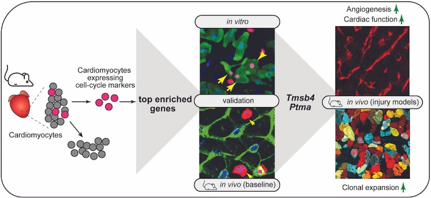
“The adult mammalian heart is a post-mitotic organ. Even in response to necrotic injuries, where regeneration would be essential to reinstate cardiac structure and function, only a minor percentage of cardiomyocytes undergo cytokinesis… In this study, we aimed to determine the gene expression profile of proliferating adult cardiomyocytes in the mammalian heart after myocardial ischaemia, to identify factors to can promote cardiac regeneration… Here, we demonstrate increased 5-ethynyl-2’deoxyuridine incorporation in cardiomyocytes 3 days post-myocardial infarction in mice… Combinatorial overexpression of the enriched genes within this population in neonatal rat cardiomyocytes and mice at postnatal day 12 (P12) unveiled key genes that promoted increased cardiomyocyte proliferation. Therapeutic delivery of these gene cocktails into the myocardial wall after ischaemic injury demonstrated that a combination of thymosin beta 4 (TMSB4) and prothymosin alpha (PTMA) provide a permissive environment for cardiomyocyte proliferation and thereby attenuated cardiac dysfunction… This study reveals the transcriptional profile of proliferating cardiomyocytes in the ischaemic heart and shows that overexpression of the two identified factors, TMSB4 and PTMA, can promote cardiac regeneration.” (2)
The Role of Thymosin β4 in Cardiomyocyte Proliferation and Cardiac Regeneration
“Thymosin β4 (Tβ4) is a 43-amino acid protein that belongs to the β-thymosin family, which is highly conserved… Tβ4 has been associated with wound healing, inflammation, fibrosis, and tissue regeneration, with recent studies suggesting that Tβ4 can help prevent inflammation and fibrosis in the eye, skin, lung, and liver… Tβ4 is a potent protective factor that can protect against myocyte damage, promote myocyte regeneration, and inhibit heart inflammation… Therefore, we propose that Tβ4 might have an antifibrotic function in the heart.” (3)